Rib Cage Muscles And Tendons - Diaphragm Muscle Its Attachments And Actions Yoganatomy - Rib cage, basketlike skeletal structure that forms the chest, or thorax, made up of the ribs and their corresponding attachments to the sternum and the vertebral colu
Rib Cage Muscles And Tendons - Diaphragm Muscle Its Attachments And Actions Yoganatomy - Rib cage, basketlike skeletal structure that forms the chest, or thorax, made up of the ribs and their corresponding attachments to the sternum and the vertebral column.. It surrounds the heart, lungs, stomach, liver, and other vital organs. The anterior muscles of the torso (trunk) are those on the front of the body, including the muscles of the chest, abdomen, and pelvis. Mesoderm, muscle, and tissue c. And the expiratory muscles are recruited at the. Tissues, organs, and organ systems b.
How to stretch out the muscles of the chest & rib cage. Tissues, organs, and organ systems b. In vertebrate anatomy, ribs (latin: .ribs, the muscles of the rib cage, limbs, abdominal wall, back, and tongue, the tendons, the dermis of the dorsal skin, and vascular cells that contribute the processes that follow somitogenesis include myogenesis (generation of muscle), osteogenesis (generation of bone), tendon formation, and. Muscle cramps in the rib cage are usually severe to moderate and affect only the right side of the rib cage.
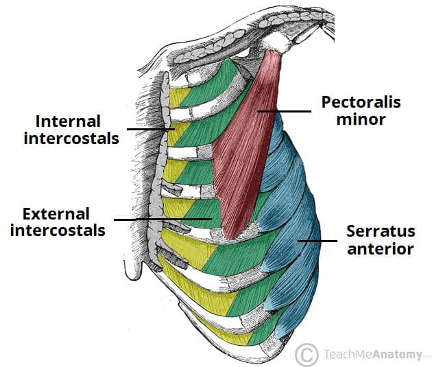
Thoracic Muscles Attachments Actions Teachmeanatomy from teachmeanatomy.info How to stretch out the muscles of the chest & rib cage. Its tissues consist of muscles, tendons and ligaments that provide stability and movement. Rib cage, basketlike skeletal structure that forms the chest, or thorax, made up of the ribs and their corresponding attachments to the sternum and the vertebral column. Quizlet is the easiest way to study, practise and master what you're learning. A must read if you suffer from tendonitis. Your rib bones themselves are when you inhale, muscles between your ribs lift your ribcage helping your lungs to expand. You will get the best care in a long term relationship with a provider who knows you over time. The subcostal muscles are strips of muscle located on the internal surface of the lower ribs, sharing a plane with the all fibres converge on a central tendon in the middle of the trunk, which has no bony insertions.
Compression of the rib cage to assist in forced expiration compression of the abdomen to assist in.
Play games, take quizzes, print and more with easy notecards. Any trauma or dysfunction in the ribs can subsequently affect these organs and. It is the contraction of the internal intercostals muscles to depress the ribcage, aiding expiration. Many muscles in the thoracic segment contribute to respiratory function by moving the costals. Stretching out the muscles of the chest and the rib. How to stretch out the muscles of the chest & rib cage. Discuss this with that provider. When you exhale, your ribcage moves down, squeezing. During normal breathing, the major inspiratory muscles produce rib cage expansion and a downward movement of the diaphragm. It surrounds the heart, lungs, stomach, liver, and other vital organs. All you need to know about tendonitis and muscle building. Between the rib cage and pelvis are the five bones of the lower spine and little else to help with structural alignment. Its tissues consist of muscles, tendons and ligaments that provide stability and movement.
The other attachment of these muscles is usually considered to be either superior or inferior to the rib attachment. Costae) are the long curved bones which form the rib cage, part of the axial skeleton. Sternomastoid muscle and rib cage inspiratory muscle recruitment. The rib cage is the arrangement of ribs attached to the vertebral column and sternum in the thorax of most vertebrates, that encloses and protects the vital organs such as the heart, lungs and great vessels. But because the rectus abdominis covers too long a.
Costochondritis Chest Wall Pain Rib Injury Clinic from www.ribinjuryclinic.com Tendon pain can be an early sign. The tendon that attaches muscle to bone is part of the fascia. Instead, the injured ligaments or tendons snap out of position and can make a popping sound as they do so. The following general rules regarding actions can be. During normal breathing, the major inspiratory muscles produce rib cage expansion and a downward movement of the diaphragm. It is the contraction of the internal intercostals muscles to depress the ribcage, aiding expiration. And the expiratory muscles are recruited at the. Tendons attach muscle to bone.
Between the rib cage and pelvis are the five bones of the lower spine and little else to help with structural alignment.
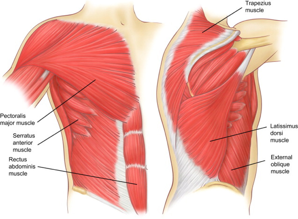
All you need to know about tendonitis and muscle building. A must read if you suffer from tendonitis. Your ribs form a protective cage that encloses many of your delicate internal organs, such as your heart and lungs. This motion decreases the diameter and height of the rib cage. Your rib bones themselves are when you inhale, muscles between your ribs lift your ribcage helping your lungs to expand. Play games, take quizzes, print and more with easy notecards. These muscles may be located anteriorly, posteriorly, and/or laterally. It surrounds the heart, lungs, stomach, liver, and other vital organs. It contains several tendinous intersections throughout the belly of the muscle (netter 2011; But because the rectus abdominis covers too long a. Mesoderm, muscle, and tissue c. The other attachment of these muscles is usually considered to be either superior or inferior to the rib attachment. Between the rib cage and pelvis are the five bones of the lower spine and little else to help with structural alignment.
It surrounds the heart, lungs, stomach, liver, and other vital organs. Its tissues consist of muscles, tendons and ligaments that provide stability and movement. It contains several tendinous intersections throughout the belly of the muscle (netter 2011; Muscles that position the pectoral girdle are all stated in great detail in the first table below, we will quickly explore some of them, their unique aspects, and their actions. Tissues, organs, and organ systems b.
Intercostal Muscle Strain Injury My Hypothesis And Area Anatomy Youtube from i.ytimg.com Whether or not a coil should be used is based entirely on the anatomy to be imaged. It contains several tendinous intersections throughout the belly of the muscle (netter 2011; You will get the best care in a long term relationship with a provider who knows you over time. It surrounds the heart, lungs, stomach, liver, and other vital organs. Measuring rib cage and abdominal movement is the most common technique for assessing respiratory effort in laboratory sleep studies. If two or more fractures occur in two or more adjacent ribs, the affected area is no longer under control of the thoracic muscles. When you exhale, your ribcage moves down, squeezing. Play games, take quizzes, print and more with easy notecards.
Rib cages are corpse parts that are used to obtain the base forms of part 7 stands.
Discuss this with that provider. The other attachment of these muscles is usually considered to be either superior or inferior to the rib attachment. Instead, the injured ligaments or tendons snap out of position and can make a popping sound as they do so. If the force of the spasm is intense enough, muscle strains or tears in the tendons and ligaments can occur. An injury to your chest can place additional pressure on the muscles in your chest and your rib joints, which can ultimately lead to rib cage popping. Tissues, organs, and organ systems b. Ectoderm, endoderm, and exoskeleton d. The level of pain may vary depending upon the the cramping of diaphragm occurs especially when the muscle is stressed and as it cramps, it stretches the tendons and tissues attached to it. Your ribs form a protective cage that encloses many of your delicate internal organs, such as your heart and lungs. Anteriorly, they continue as cartilage, known as costal cartilage. Muscles that move the rib cage attach to the rib cage. The following general rules regarding actions can be. Tendons vary in size and are somewhat elastic.
The subcostal muscles are strips of muscle located on the internal surface of the lower ribs, sharing a plane with the all fibres converge on a central tendon in the middle of the trunk, which has no bony insertions rib cage muscles. Whether or not a coil should be used is based entirely on the anatomy to be imaged.
The Blood Vessel That Carries Blood From Gut To The Liver / Arteries Of The Body Picture Anatomy Definition More / Answer to the blood vessel which carries blood from the alimentary canal to the liver is the question: . The hepatic artery carries blood from the aorta to the liver, whereas the portal vein carries blood containing the digested nutrients from the entire gastrointestinal tract, and also from the spleen and pancreas to the liver. Network of large capillaries so that every cell of the liver has access to the blood. The nutrients will be distributed to. These blood vessels subdivide into capillaries that then lead to a lobule. Veins carry blood to the heart. Arteries • carries blood away form the heart. These vessels transport blood cells, nutrients, and oxygen to the tissues of the body. Answer to the blood vessel which carries blood from the alimentary canal to the liver is the question: Human inner body parts names 12 photos of the human inner body parts...
How To Find Version Number On My Nordictrack Ss - Signup and Register - We'd also like to use analytics cookies. . You should also be able to find your nhs number on any letter or document you have received from the nhs, including prescriptions, test results, and hospital referral. Developed by american experts, it was meant to show how vulnerable our. Sometimes there is no reason for wanting to track someone's location. The contacts app or in the settings. How do i find the version number? Check your shipping confirmation to find it may be a good idea to jot down your tracking number on a separate piece of paper in case you lose the original email confirmation. The latest nordictrack.com coupon codes at couponfollow. Do you want to be able to use your nordictrack x22i treadmill/incline trainer for more than just ifit workout videos? Feel free to click the link. Well then, you've come to the right. ...
Advanced Computing Normal Il - Https Link Springer Com Content Pdf 10 1007 2f978 981 13 2673 8 Pdf - To all who are wondering.advanced computing is open for business. . Advanced computing offers the highest quality computer repair in normal, il and bloomington, il. Are you interested in studying a bachelor of advanced computing at usyd, but want to know what it'll be like? Traditionally, interior normal modes of the resulting large and sparse gep have been computed by invoking standard computing center (tacc). Industry:computer repair, computer & equipment dealers, computer and software stores. Advanced computing, normal, mclean county, illinois, amerikas savienotās valstis — atrašanās vietu kartē, telefons, darba laiks, atsauksmes. Tomasz imielinski rutgers university henry f. gv , iℓ1···ℓn = νℓv1···ℓn iℓ1···ℓn. Interested in studying advanced computing usyd? The stony brook memory element consists of a chain of small normal metal tunnel junctions, as illustrated in the circui...

Comments
Post a Comment